

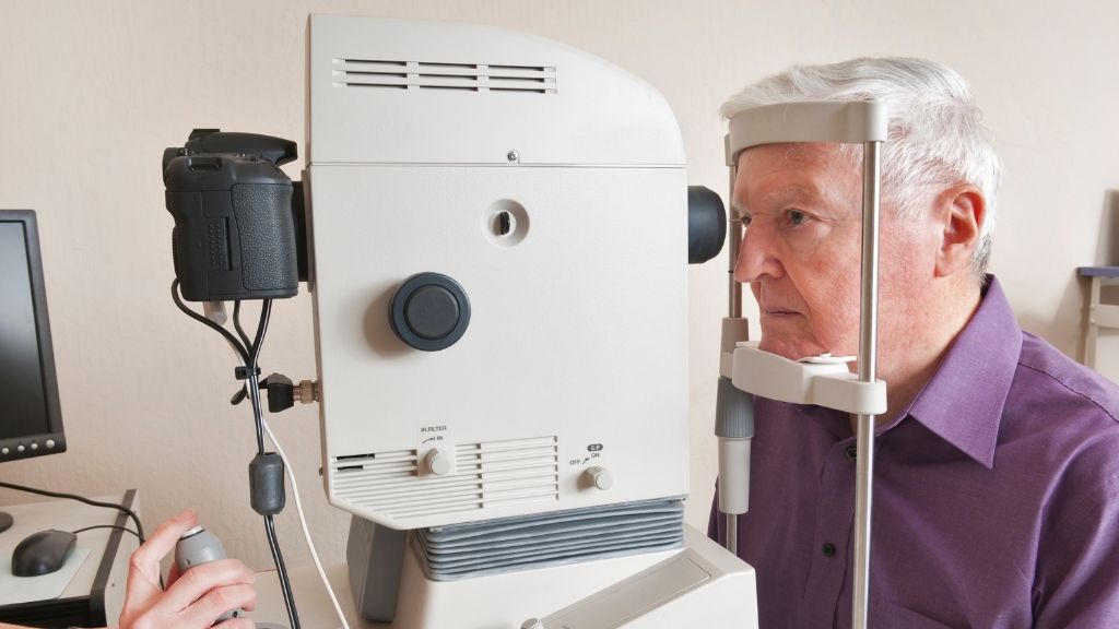
A retinal detachment occurs when the retina separates from the tissues that support it at the back of the eye. Because the retina is essential for vision, this condition requires urgent ophthalmological assessment and, in many cases, surgical treatment.
One of the most important questions for patients is how quickly retinal detachment surgery should be performed. There is no single timeframe for every case. The urgency depends on the type and extent of the detachment, the location of the retinal tear and, particularly, whether the macula, the central part of the retina responsible for detailed vision, is still attached.
This guide explains the warning signs of retinal detachment, why treatment should not be delayed, the main surgical options and what patients can expect during recovery.
Is retinal detachment an emergency?
Yes. A suspected retinal detachment requires urgent assessment by an ophthalmologist. Prompt diagnosis is important because the detachment can progress and involve the macula, potentially affecting central vision. The exact timing of treatment is determined by a retinal specialist according to each case.
What is retinal detachment?
The retina is a thin layer of light-sensitive tissue at the back of the eye that plays an essential role in vision. A retinal detachment occurs when this layer separates from its normal position. The most common form develops when a retinal tear or hole allows fluid to pass underneath the retina.
Other forms can occur when scar tissue pulls on the retina or when fluid accumulates beneath it without a retinal tear.
What are the symptoms of retinal detachment?
Retinal detachment symptoms are usually painless and can appear suddenly. Recognising them is particularly important because central vision may initially remain relatively normal.
Sudden floaters
A sudden increase in dots, lines or cobweb-like shapes moving across the field of vision.
Flashes of light
Brief flashes, often noticed at the side of the visual field, particularly in dim conditions.
Shadow or curtain
A dark area or curtain-like shadow that appears to move across part of the visual field.
Loss of peripheral vision
Part of the side vision may disappear as the detached area becomes larger.
Blurred vision
Vision may become blurred or distorted, particularly if the central retina becomes involved.
Sudden visual loss
A significant decrease in vision requires immediate ophthalmological assessment.
How quickly should retinal detachment surgery be performed?
The timing of retinal detachment surgery is determined individually. One of the most important considerations is whether the macula is still attached.
If the macula remains attached, treatment may be particularly time-sensitive because one of the objectives is to repair the retina before the detachment progresses towards the area responsible for central, detailed vision.
If the macula has already detached, surgery is still generally required promptly. The retinal specialist considers factors such as how long the macula may have been detached, the location and extent of the detachment, the number of retinal breaks and the overall condition of the eye.
How long can retinal detachment go untreated?
There is no universally safe period for leaving a diagnosed retinal detachment untreated. Some detachments can progress rapidly. Patients should not wait for central vision to deteriorate before seeking medical attention. The appropriate timing of treatment should be established by a retinal specialist.
Retinal tear vs retinal detachment: are they the same?
A retinal tear is a break in the retina. If fluid passes through that break and accumulates underneath, the retina can detach. Certain retinal tears detected before significant detachment develops may be treated with laser photocoagulation or cryotherapy. Once a significant retinal detachment has developed, surgery is commonly required to reposition the retina.
How is retinal detachment treated?
Retinal detachment treatment depends on the type of detachment, the location and number of retinal breaks, the condition of the vitreous and other characteristics of the eye.
Vitrectomy
The vitreous gel is removed to relieve traction on the retina. Retinal breaks are treated and a gas bubble or silicone oil may be used to support healing.
Scleral buckle
A silicone band or element is placed on the outside of the eye to help close the retinal break and support reattachment.
Pneumatic retinopexy
In selected cases, a gas bubble is injected into the eye to push the retina into position while the retinal break is sealed with laser or cryotherapy.
Vitrectomy for retinal detachment
Vitrectomy is frequently used to repair retinal detachment. The surgeon removes the vitreous gel, releases traction affecting the retina and treats the retinal tears. A gas bubble or silicone oil may then be used to support the retina during healing.
If a gas bubble is used, specific head positioning may be required. Air travel is generally prohibited while intraocular gas remains in the eye because changes in atmospheric pressure can cause the bubble to expand dangerously.
Scleral buckle surgery
Scleral buckle surgery uses a silicone element placed on the outside of the eye to reduce traction and help close the retinal break. In selected or more complex cases, it may be combined with vitrectomy.
How is the surgical technique selected?
Location and extent
The position and size of the detachment and whether the macula is involved influence treatment planning.
Number of retinal tears
Multiple or difficult-to-access breaks may require a different surgical strategy.
Condition of the vitreous
Vitreous traction can influence whether vitrectomy is appropriate.
Previous eye surgery
Previous cataract or retinal procedures may affect surgical planning.
What is recovery like after retinal detachment surgery?
Recovery after retinal detachment surgery varies considerably. The retina may be successfully reattached while vision continues to improve gradually over weeks or months. Patients may initially experience blurred vision, mild discomfort or redness, and eye drops are commonly prescribed.
When gas has been placed inside the eye, a particular head position may be required. Patients should follow their surgeon’s instructions and confirm when they can safely resume driving, exercise, lifting and travel.
When will vision return after retinal detachment surgery?
Visual recovery depends particularly on whether the macula was detached and how long it had been affected. Vision may improve gradually as the eye heals, although some patients can have persistent changes in visual acuity or distortion even after successful retinal reattachment.
Retinal detachment surgery in Barcelona
Barcelona International Hospitals brings together hospitals with specialist ophthalmology services for the diagnosis and treatment of complex retinal conditions. Access to retina specialists, advanced imaging and vitreoretinal surgery can be particularly important when treatment is time-sensitive.
Hospitals within the Barcelona International Hospitals network providing specialised ophthalmological care include:
barnaclínic+ (Hospital Clínic Group)
Provides access to specialised ophthalmological diagnosis and surgical treatment within the Hospital Clínic Barcelona Group.
Provides specialist care for retinal and vitreous conditions, including surgical treatment for retinal detachment.
Provides specialised ophthalmological care for retinal and macular disorders, supported by advanced diagnostic and surgical technology.
Hospital Quirónsalud Barcelona
Provides specialised ophthalmological care with access to advanced diagnostic and surgical facilities. Ophthalmology is also represented among the BIH specialists recognised among Spain’s leading doctors.
International patients seeking retinal treatment in Barcelona
Barcelona International Hospitals can help international patients identify an appropriate hospital and specialist team according to their clinical needs. Patients considering medical care in the city can also consult our guide to private hospitals in Barcelona for international patients. However, a suspected retinal detachment is time-sensitive. Patients experiencing sudden flashes, new floaters, loss of peripheral vision or a curtain-like shadow should seek urgent ophthalmological assessment rather than delay care while organising international travel.
When transfer to Barcelona is clinically appropriate, previous ophthalmology reports and retinal imaging can help the receiving team evaluate the case. After surgery, patients with an intraocular gas bubble must not fly until their ophthalmologist confirms that air travel is safe.
This article is for informational purposes only and does not replace professional medical advice, diagnosis or treatment. If you experience symptoms that may indicate retinal detachment, seek urgent ophthalmological assessment.